Kidney Stones Lateral X Ray

Abdominal Radiology Quiz14 Radiology Radiography Abdominal

Image Result For Where Is The Kidney Ureter And Bladder In Relation To Left Transverse Processes Of L4 Bladder Relatable Pelvis

Bladder Calculus Radiology Case Radiopaedia Org Radiology Radiology Imaging Radiography

Pin On Gastrointestinal Radiology
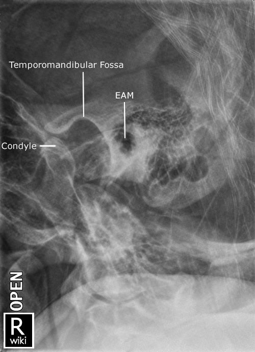
Pin By Jeremy Enfinger On Radiographic Anatomy Radiology Imaging Radiology Schools Medical Knowledge

Mobile Abdominal X Ray Medical Radiography Medical Anatomy Medical Knowledge

When the stone is not on the focus the sound waves can damage the soft tissue of the kidney.
Kidney stones lateral x ray. This x ray using contrast reveals a kidney stone at the junction of the kidney and the tube that connects the kidney to the bladder ureter. Kidney stone disease also known as nephrolithiasis or urolithiasis is when a solid piece of material kidney stone develops in the urinary tract. Upon his return to taichung he went for a medical examination where blood studies and x rays gave.
This will help them to work out a treatment plan. It was acid in reaction and contained a small amount of albumen. Doctors can use it to help them diagnose.
Abdominal x rays can show the location of kidney stones in the urinary tract. Microscopic examination disclosed 25 red blood cells in a low power field and 12 pus cells. X rays including an intravenous pyelogram ivp where dye is injected into the bloodstream before the x ray is taken.
A small stone may pass without causing symptoms. This damage can be prevented by a feedback mechanism that determines the place of kidney stones depending on the images taken from eswl device. If a stone grows to more than 5 millimeters 0 2 in it can cause blockage of the ureter resulting in severe pain.
X ray image of kidney stone. The x ray technician will position the x ray machine over or in front of your abdomen and ask you to hold your breath so the picture won t be blurry. Kidney stones typically form in the kidney and leave the body in the urine stream.
The specific gravity of the urine was 1 029. The x ray technician then may ask you to change position for additional pictures. Most kidney stones will pass by themselves within three to six weeks.

Kidney Stone Kidney Stones Funny X Ray Kidney Stones
Medullary Nephrocalcinosis Upper Photo Of Conventional Radiograph Of Abdomen And Lower Photo Of Coro Radiology Imaging Radiology Diagnostic Medical Sonography

Abdominal Radiographic Anatomy Wikiradiography Radiology Imaging Medical Radiography Radiology Schools

Pin On Because People Matter

Medullary Nephrocalcinosis Upper Photo Of Conventional Radiograph Of Abdomen And Lower Photo Of Coro Radiology Imaging Radiology Diagnostic Medical Sonography

Untitled Document Radiology Imaging Medical Knowledge Radiology Student

There Are Stones And Then There Are Stones This Is A Gigantic Ureteric Stone See The Complete Case Http Radiopaedia Org Cases Radiology Cool Tools Stone

Pin On Ct Scans

Rosh Review Medical School Stuff Medical Mnemonics Large Bowel

Pin By Kathy I On Radiology Radiography Radiology Medical

The Cobra Head Sign Or Spring Onion Sign Refers To Dilatation Of The Distal Ureter Surrounded By A Thin Lucent Radiology Medical Imaging Science And Nature

Autosomal Dominant Polycystic Kidney Disease Radiology Case Radiopaedia Org Polycystic Kidney Disease Renal Disease Kidney Disease

Untitled Document Gallstones X Ray
